


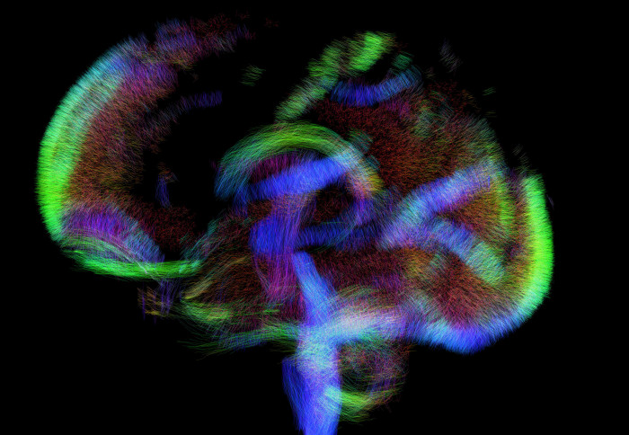
A baby's brain at 33 weeks
Global S&T Development Trend Analysis Platform of Resources and Environment
| 800 newborn brain scans made available to help clarify how some diseases develop | |
| admin | |
| 2021-06-02 | |
| 发布年 | 2021 |
| 语种 | 英语 |
| 国家 | 英国 |
| 领域 | 资源环境 |
| 正文(英文) |
A baby's brain at 33 weeks 
The Developing Human Connectome Project (dHCP) has made the scans of 800 baby brains available to study how developmental disorders progress. The scans, part of a large-scale open-source project, are available online for researchers who study brain development in foetuses and babies. The research team hope the scans will provide clues on how conditions like autism develop, and how problems like air pollution in pregnancy affect brain growth. They also want to clarify how brain wiring and function develops during pregnancy and after birth. The dHCP dataset presents a unique resource for understanding brain development using advanced computational tools. Professor Daniel Rueckert Department of Computing The dHCP is a collaboration between King’s College London, Imperial College London and the University of Oxford, and the images are the team's third open-source large-scale data release of this project. The researchers are currently analysing the images to ask questions about how the brain develops and whether early birth negatively affects brain development. They are also looking into using artificial intelligence (AI) to answer these questions. Co-investigator Professor Daniel Rueckert from Imperial’s Department of Computing said: “The dHCP dataset presents a unique resource for understanding brain development using advanced computational tools. We have already started exploring the development of AI approaches to ask questions about how the brain develops.” Principal Investigator Professor David Edwards from King’s said there have never been this many brain scans of babies put out for researchers to use. He said: “It’s completely novel in the sense that it’s the largest data set yet created and it’s free for people to use. We’re providing the world with a chance to make a map of whatever feature of normal development they want to study – whether it’s the growth of the frontal lobe, or cerebellar function – whatever it is, this is the data you can use to make your map.” These new scans complete a neonatal dataset of brain images and associated clinical data like sex, age at birth, age at scan, birthweight, head circumference and radiology score. The imaging data includes structural imaging to measure and quantify different structures in the brain, structural connectivity data (diffusion MRI) to explore how different brain regions are connected, and functional connectivity data (resting-state fMRI) to measure how different brain regions communicate. We hope that the resulting data will be valuable to many researchers and will provide an enduring resource that will help significantly to drive forward understanding of human brain development. Professor Jo Hajnal King's College London For these scans, most of the babies were imaged while naturally asleep. If the baby woke up, scanning was stopped and attempts made to resettle before proceeding. If a baby moved, the team ensured all the data was motion corrected, largely using methods developed specifically for the dHCP project, in order to produce highly detailed and rich information on brain development. Co-investigator Professor Jo Hajnal from King’s said: “During the project we have developed dedicated receiver coils and patient handling, as well as MR acquisition, image reconstruction and analysis methods all specifically optimised for neonates. We hope that the resulting data will be valuable to many researchers and will provide an enduring resource that will help significantly to drive forward understanding of human brain development.” Co-investigator Professor Steve Smith from the University of Oxford said: "This is the result of many years hard coordinated work across our 3 universities, and has resulted in the most amazing, rich data on how the brain develops just before and after birth. We're very excited to release the dataset to the international research community.” Future data releases will provide foetal images (imaging babies in the womb), more detailed ancillary data as well as genetic and clinical data. The research consortium is funded by a €15 million Synergy grant from the European Research Council. The aim is to ensure data is shared as widely as possible. Future releases will include data from babies still in the womb as well as information on children’s genes and their abilities as they grow up. Researchers can now download the data pack. Image and GIFs: dHCP |
| URL | 查看原文 |
| 来源平台 | Imperial College London |
| 文献类型 | 新闻 |
| 条目标识符 | http://119.78.100.173/C666/handle/2XK7JSWQ/328633 |
| 专题 | 资源环境科学 |
| 推荐引用方式 GB/T 7714 | admin. 800 newborn brain scans made available to help clarify how some diseases develop. 2021. |
| 条目包含的文件 | 条目无相关文件。 | |||||
| 个性服务 |
| 推荐该条目 |
| 保存到收藏夹 |
| 查看访问统计 |
| 导出为Endnote文件 |
| 谷歌学术 |
| 谷歌学术中相似的文章 |
| [admin]的文章 |
| 百度学术 |
| 百度学术中相似的文章 |
| [admin]的文章 |
| 必应学术 |
| 必应学术中相似的文章 |
| [admin]的文章 |
| 相关权益政策 |
| 暂无数据 |
| 收藏/分享 |
除非特别说明,本系统中所有内容都受版权保护,并保留所有权利。
修改评论