Global S&T Development Trend Analysis Platform of Resources and Environment
| Vascular development : finding the right itinerary | |
| admin | |
| 2018-12-21 | |
| 发布年 | 2018 |
| 语种 | 英语 |
| 国家 | 法国 |
| 领域 | 地球科学 |
| 正文(英文) | Researchers at the CNRS, Université Paris Diderot and Université Paris Descartes have demonstrated how the growth of veins and arteries unfolds in embryonic development. Their findings prove that, contrary to prevailing opinion, blood exits arteries at an upstream point of the vessel, not its end. Additionally, veins develop in an interlaced pattern between arteries, where blood passes through the capillaries. Results appear in the December 21, 2018 edition of Communications Biology.
Nutrients and oxygen are delivered to organs by the blood which moves through them in tiny vessels called capillaries: fresh blood arrives through arteries and exits, carrying metabolic waste, through veins. The correct arrangement of this vascular system, or vasculature, is therefore essential in embryonic development. Researchers have long observed that as an organ grows, arteries and veins naturally interlace in a spectacular manner that leaves between them a small network of capillaries arranged in a perfectly spaced out pattern. How do these vessels 'find their way'?
Using a new imaging technique (patent pending), teams at the Laboratoire Matière et Systèmes Complexes (CNRS/Université Paris Diderot) and the Unité de technologies chimiques et biologiques pour la santé (CNRS/INSERM/Université Paris Descartes/Chimie Paristech) have studied with precision the development of vasculature and vessels via a standard model in vascular biology research. One unexpected finding contradicted the long-held belief that arteries are cylindrical. To the contrary, their extremities are flattened and attached to the tissue, like a crushed hose. Likewise, blood flow is nearly non-existent is this part of the artery. Instead, blood exits the artery via capillaries located prior to the flattened end and then enters the veins. (See photo) Guided by the flow of blood, veins grow in an interlaced manner between the arteries, rather than against the artery ends, where blood does not flow. Thanks to this phenomenon, arteries and veins develop alternately, with the ends of veins level with the irrigated capillaries, upstream of artery ends. The research team will now focus its attention on the development of dysfunctional vascular structures such as those which appear in tumours caused by certain cancers. 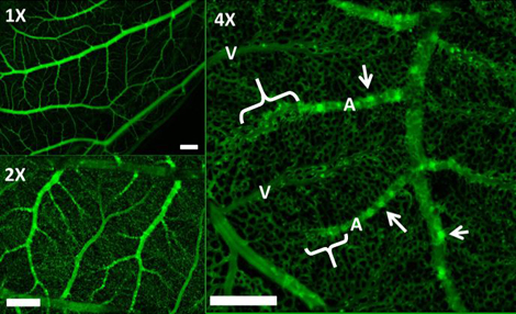 © Vincent Fleury/Nature Successive enlargements of a vascular structure showing interlacing of arteries (A) and veins (V). Arrows point to blood flow exit points toward capillaries, upstream of artery ends. Brackets indicate the flattened ends of arteries. Download the press release :  Bibliography:Direct imaging of capillaries reveals the mechanism of arteriovenous interlacing in the chick chorioallantoic membrane. Sophie Richard, Amanda Brun, Antonio Tedesco, Benjamin Gallois, Naoual Taghi, Philippe Dantan, Johanne Seguin and Vincent Fleury. Communications Biology, December 21, 2018. DOI : 10.1038/s42003-018-0229-x Contacts: CNRS researcher | Vincent Fleury | T +33 1 57 27 62 56 | vincent.fleury@univ-paris-diderot.fr |
| URL | 查看原文 |
| 来源平台 | Centre national de la recherche scientifique |
| 文献类型 | 新闻 |
| 条目标识符 | http://119.78.100.173/C666/handle/2XK7JSWQ/107030 |
| 专题 | 地球科学 |
| 推荐引用方式 GB/T 7714 | admin. Vascular development : finding the right itinerary. 2018. |
| 条目包含的文件 | 条目无相关文件。 | |||||
| 个性服务 |
| 推荐该条目 |
| 保存到收藏夹 |
| 查看访问统计 |
| 导出为Endnote文件 |
| 谷歌学术 |
| 谷歌学术中相似的文章 |
| [admin]的文章 |
| 百度学术 |
| 百度学术中相似的文章 |
| [admin]的文章 |
| 必应学术 |
| 必应学术中相似的文章 |
| [admin]的文章 |
| 相关权益政策 |
| 暂无数据 |
| 收藏/分享 |
除非特别说明,本系统中所有内容都受版权保护,并保留所有权利。


修改评论